생체조직의 3차원 사진 촬영
페이지 정보
불만이 작성일2003-06-10 10:09관련링크
본문
Nature 6월 10일자
---------------------------------------------------
레이저를 이용한 3차원 생체조직 사진 촬영
레이저를 이용한 생체 조직의 3차원 촬영 방법이 계발되었다. 기존의 방법은 조직을 자르고 염색하여 현미경으로 관찰하였으나 레이저를 이용한 새로운 기술은 이를 대체할 수 있을 것으로 보인다. Colorado School of Mines 의 Jeff Squier 가 계발한 이 기술은 적외선 레이저를 형광 염료 처리한 조직 샘플에 조사하였을 때 염료 분자가 에너지를 얻었다가 얻은 에너지를 빛으로 방출하는 것을 측정 하는 방법이다. 이러한 방법을 이용하여 수 시간 만에 복잡한 실험실 작업을 거치지 않고 조직 시료의 디지털 화상 자료를 얻을 수 있었다. 실험팀은 이 방법을 쥐의 뇌에 대하여 적용하여 3차원 이미지를 얻는데 성공하였다. 또한 쥐의 배아에 대해서도 적용하였는데, 기존의 방법으로는 이러한 연한 조직을 다룰 수 없었다.
Lasers take 3D brain scans
Infrared light probes tissue a slice at a time.
10 June 2003
GEOFF BRUMFIEL
A new technique for probing tissue samples with lasers could give researchers easy access to three-dimensional images of brain samples and pieces of other organs.
The system could replace the current methods of imaging soft tissue, says one of its developers Jeff Squier, a physicist at the Colorado School of Mines. Typically, samples are frozen, sliced and dyed, before being examined under the microscope. This process can distort key details.
Squier's team places a fresh, dice-sized sample of rat brain tissue in front of a high-powered laser. The laser emits short, bright pulses of infrared light that stimulate fluorescent dyes that are either genetically engineered or manually brushed into the sample. The dye then emits light at a different wavelength that is picked up by detectors around the tissue.
The laser sweeps through the sample, taking a digitized image of its surface. Then a second laser burns away scanned tissue and the process begins anew. When the whole sample has been scanned, all that remains is a digital, three-dimensional picture of the chunk of rat brain. "The sample goes from brains to bits," he says.
Squier presented the first results from the device at last week's Conference on Lasers and Electro-Optics in Baltimore, Maryland. He developed it in collaboration with researchers at the University of California, San Diego and at Science Applications International Corporation in Arlington, Virginia.
Ultimately, laser scanning could automatically create digital images in just hours without lab technicians having to painstakingly freeze, slice, stain and set samples. A similar system is also showing promise in the imaging of rat embryos. Traditional techniques can't handle such soft tissue.
Geoff Brumfiel is Washington Physical Sciences Correspondent for the journal Nature

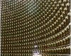
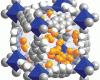
Lasers turn a sample from brains to bits.
© J. Squier
---------------------------------------------------
레이저를 이용한 3차원 생체조직 사진 촬영
레이저를 이용한 생체 조직의 3차원 촬영 방법이 계발되었다. 기존의 방법은 조직을 자르고 염색하여 현미경으로 관찰하였으나 레이저를 이용한 새로운 기술은 이를 대체할 수 있을 것으로 보인다. Colorado School of Mines 의 Jeff Squier 가 계발한 이 기술은 적외선 레이저를 형광 염료 처리한 조직 샘플에 조사하였을 때 염료 분자가 에너지를 얻었다가 얻은 에너지를 빛으로 방출하는 것을 측정 하는 방법이다. 이러한 방법을 이용하여 수 시간 만에 복잡한 실험실 작업을 거치지 않고 조직 시료의 디지털 화상 자료를 얻을 수 있었다. 실험팀은 이 방법을 쥐의 뇌에 대하여 적용하여 3차원 이미지를 얻는데 성공하였다. 또한 쥐의 배아에 대해서도 적용하였는데, 기존의 방법으로는 이러한 연한 조직을 다룰 수 없었다.
Lasers take 3D brain scans
Infrared light probes tissue a slice at a time.
10 June 2003
GEOFF BRUMFIEL
A new technique for probing tissue samples with lasers could give researchers easy access to three-dimensional images of brain samples and pieces of other organs.
The system could replace the current methods of imaging soft tissue, says one of its developers Jeff Squier, a physicist at the Colorado School of Mines. Typically, samples are frozen, sliced and dyed, before being examined under the microscope. This process can distort key details.
Squier's team places a fresh, dice-sized sample of rat brain tissue in front of a high-powered laser. The laser emits short, bright pulses of infrared light that stimulate fluorescent dyes that are either genetically engineered or manually brushed into the sample. The dye then emits light at a different wavelength that is picked up by detectors around the tissue.
The laser sweeps through the sample, taking a digitized image of its surface. Then a second laser burns away scanned tissue and the process begins anew. When the whole sample has been scanned, all that remains is a digital, three-dimensional picture of the chunk of rat brain. "The sample goes from brains to bits," he says.
Squier presented the first results from the device at last week's Conference on Lasers and Electro-Optics in Baltimore, Maryland. He developed it in collaboration with researchers at the University of California, San Diego and at Science Applications International Corporation in Arlington, Virginia.
Ultimately, laser scanning could automatically create digital images in just hours without lab technicians having to painstakingly freeze, slice, stain and set samples. A similar system is also showing promise in the imaging of rat embryos. Traditional techniques can't handle such soft tissue.
Geoff Brumfiel is Washington Physical Sciences Correspondent for the journal Nature
Lasers turn a sample from brains to bits.
© J. Squier
댓글 0
등록된 댓글이 없습니다.
- 이전
- 플라즈마 벽
- 다음
- This Week in Science